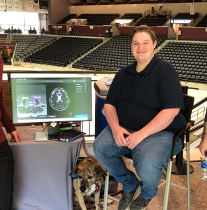
Luka Woodson, an EAC student researcher, recently won 1st Place in Interdisciplinary at the undergraduate level at the 2023 UA Little Rock Research & Creative Works Expo for his project entitled ‘Towards a more natural interaction for medical imaging annotation tools’. Woodson received a cash prize and a certificate. Woodson also received a 1st Place certificate from Donaghey College of Science, Technology, Engineering, and Mathematics for the Research Expo.
The Medical Imaging Annotation Tool is a game-changing technology created with medical professionals and researchers in mind. This cutting-edge application is intended to make X-ray image annotation easier by using bounding boxes, polygons, and labels with unparalleled simplicity. Say goodbye to complex software and steep learning curves with the Medical Imaging Annotation Tool, as its easy interface enables users to quickly discover and transmit critical information about a patient’s health, increasing workflow efficiency and saving vital time.
The Medical Imaging Annotation Tool’s compatibility with industry-standard file formats is one of its most notable advantages. This application provides smooth interaction with a wide range of image files by supporting well-known file types such as the MS COCO annotation format and DICOS. Whether using current datasets or adding other resources, the Medical Imaging Annotation Tool ensures unsurpassed compatibility and flexibility, increasing productivity and extending possibilities.
The incorporation of the innovative dual viewport capability within the Medical Imaging Annotation Tool is an exceptional advantage. This novel feature provides users with not one, but two viewports simultaneously, allowing them to modify and compare two photos simultaneously. This feature’s value is its capacity to assist in efficient analysis, allowing for rapid comparisons and detailed assessments. By visualizing and interpreting images within a single interface, healthcare professionals and researchers gain unparalleled convenience and precision, revolutionizing their approach to image analysis.
Recognizing the challenges associated with existing software, the Medical Imaging Annotation Tool’s developers have painstakingly designed a powerful and user-friendly solution. Embrace a new era of efficiency and precision in medical imaging analysis. Join the transformative movement in how X-ray images are handled, enabling healthcare professionals to focus on their primary objective: providing outstanding care to their patients.
Try the Medical Imaging Annotation Tool today to experience the future of medical image annotation. Discover how its seamless file compatibility and dual viewport features can enhance your workflow, increase your capabilities, and open new horizons in medical image analysis.


